Archives of Preventive Medicine
Analysis of current and future technologies of capsule endoscopy: A mini review
Alexander P Brown and Ahalapitiya H Jayatissa*
Cite this as
Brown AP, Jayatissa AH (2020) Analysis of current and future technologies of capsule endoscopy: A mini review. Arch Prev Med 5(1): 031-034. DOI: 10.17352/apm.000016Many existing methods of endoscopy can be very uncomfortable and potentially even painful for a patient. Using a conventional endoscope is also limited in its usable range, unable to access a majority of the small bowel. Recent advancements in LEDs, optical design, and MEMS (microelectromechanical systems) technologies have provided the ability to create a wireless endoscope. Since its inception, the capsule endoscope has seen advancements in existing technology as well as the introduction of new components. As the capsule endoscope continues to advance, more application possibilities will grow as well.
Introduction
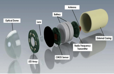
Endoscopy is a procedure in which a long flexible tube equipped with a camera is used to enter the human body to investigate unusual symptoms experienced in the esophagus, stomach, small portions of the small intestine, and the colon. This procedure can be very invasive, as it involves the insertion of an endoscope through a patient’s mouth or anus [1,2]. The nature of the endoscope also lends to the increased chance for damage or lesions in the subjected organs [3]. The development and application of the capsule endoscope has solved many of the common issues seen in the conventional endoscope. This digestible camera is the primary medical device used in a procedure known as capsule endoscope is a small wireless device capable of imaging the gastrointestinal tract of a patient Figure 1 [3].
The capsule endoscope packages many of the capabilities of the conventional endoscope and places it into a swallowable pill form [1]. The first capsule endoscope device was introduced in 2000 by an Israeli company known as Given Imaging. Given Imaging originally named their endoscope “M2A” or mouth to anus, but later changed it to “PillCam™” [4,5]. The capsule device contains several components including, an external capsule, a transparent optical window, LEDs (light-emitting diodes), a lens, an image sensor, batteries, a radio frequency transmitter, and an antenna. The receiver carried by the patient collects data transmitted from pill-cam. The information is then processed and stored using a computer with specialized software [5,6].
Capsule endoscope components
A typical capsule endoscope contains several primary components; the external casing, optical window, LED array, optical lens, CMOS image sensor, radio frequency transmitter, antenna, and a power source. The following is a general analysis of the purpose of each component in the assembly of a capsule endoscope.
The outermost portion of the endoscope is the external casing and the optical window. The optical window allows for light from the integrated LEDs to illuminate the local environment. The outer casing protects the electrical components from contacting any potentially damaging bodily fluids. Inside the capsule, an array of LEDs is placed around the camera lens [5]. The number of LEDs vary by model but is commonly between 4-8. The first component of the imaging system is the optical lens. The lens is the means by which light is focused onto the image sensor. The capsule endoscope utilizes a short focal length (the distance between the center of the lens and the image sensor), which provides a wide angle of view [1].
The image sensors most commonly used in capsule endoscopes are CMOS (complementary metal oxide semiconductor) sensors. CMOS sensors can contain several electronic components on one chip, making them a preferred choice. The ability of a CMOS chip to contain functions such as exposure and timing controls on the same chip makes it particularly advantageous over the CCD (charge-coupled device) sensor, which requires several individual chips to achieve the same functionality [7,8]. The CMOS sensors’ integrated exposure control allows it to better adapt to a low light environment. The CMOS’s low power consumption also helps make it the superior choice for this application [6,7] Figure 2.
A radio-frequency transmitter and antenna transmit data out of the endoscope. The combination of the RF transmitter and antenna enables communication between the endoscope and the receiver. The receiver is a device worn around the patient’s waist and is required to collect the images, as the endoscope does not have onboard storage [1]. Two silver oxide batteries power the electrical systems of the capsule endoscope [5]. The smaller the capsule, the higher the overall patient comfort. The size of the internal components, however, forces certain limitations. All currently available PillCam™ capsules have an outer diameter of 11mm. Differences exist in the overall length, varying between 26mm and 32mm [9,11]”.
Capsule endoscope characteristics
Several different characteristics define the performance of a capsule endoscope; capsule size, image quality (resolution), field of view (viewing angle), frame rate, and battery life.
The quality of an image can heavily influence a doctor’s ability to make a proper diagnosis. The first iteration of the PillCam™ SB (small bowel) had an image sensor resolution of 256x256 pixels [12]. This resolution remained the standard for all PillCam™ capsules until the latest iteration of the SB, the SB3. The SB3 increased the resolution to 340x340 pixels [13]. Competing capsule endoscopes have also used various sensor resolutions, such as the ENDOCAPSULE from Olympus with a resolution of 512x512, or the MiroCam® from IntroMedic with a resolution of 320x320 pixels [12,14].
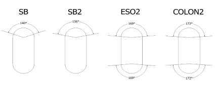
The viewing angle of an endoscope is determined by how much light passes through the lens [15]. A wider viewing angle offers a greater area of observation [1]. The original PillCam™ SB had a viewing angle of 140 degrees. The viewing angle was increased in later models (SB2 & SB3) to 156 degrees [12,13].
Communication between the capsule and the receiver is vital for data collection. The lack of any means of data storage inside the capsule endoscope means that the information must be transmitted out. The combination of the RF transmitter and antenna make this possible. The transmitter functions at roughly 432 MHz, continually sending outward to communicate with the receiver [1]. The PillCam™ SB, with a frame rate of 2 fps and battery life of 8 hours, would be theoretically capable of transmitting 57,600 images over the course of a procedure.
One of the most difficult challenges with the capsule endoscope is the limited power supply. The aspects that dictate the battery life include the sensor resolution, the frame rate, amount of light, and transmission frequency [16,17]. When comparing the PillCam™ SB and PillCam™ ESO from Table 1, this trade-off is apparent. Both endoscopes have a 256x256 pixel resolution CMOS Image sensor. The ESO, however, has a much higher frame rate of 14 frames per second versus the SBs 2 frames per second. The ESO also uses a dual camera set up, containing 6 LEDs per side or 12 in total. This higher frame rate and dual camera set up leads to the ESO having a battery life 24 times shorter than that of the SB.
The device utilizes several different optimization techniques to reduce power consumption. One technique uses specialized capsule endoscopes, optimized for specific applications. The PillCam™ ESO, for example, is used exclusively to view the esophagus. The brief travel time and immediate entry into the esophagus allow for the PillCam™ ESO to have a much shorter battery life and a much higher frame rate. Another technique used to extend battery life is the continuous switching between LEDs and the image sensor. This switching prevents the LEDs from steadily drawing power while still providing enough illumination to capture clear images [1].
Current state of the capsule endoscope
The capsule endoscope has seen increasing use by gastroenterologists since its initial release in 2001 [18]. The capsule endoscope has proven to be advantageous over the standard endoscope when it comes to patient comfort. A conventional endoscopy can be painful for a patient and often requires moderate to deep sedation. The alternative, however, can be done with the patient awake [19] Figure 3.
Current state of the capsule endoscope
The capsule endoscope has seen increasing use by gastroenterologists since its initial release in 2001 [18]. The capsule endoscope has proven to be advantageous over the standard endoscope when it comes to patient comfort. A conventional endoscopy can be painful for a patient and often requires moderate to deep sedation. The alternative, however, can be done with the patient awake [19]. Conventional endoscopes are also limited in their usable range, usually able to visualize only a small portion of the small bowel. The capsule endoscope, however, is capable of imaging the entire gastrointestinal tract [19]. Although many advantages exist with the capsule endoscope, it also comes with its limitations. One downside of the capsule endoscope is the possibility of retention. In under two percent of examinations, the patient retained the device [22]. Another issue with the capsule endoscope is its inability to be guided or controlled. The lack of locomotion within the device makes it difficult to investigate points of interest for extended periods of time [19].
Since the introduction of the PillCam™ SB in 2001, Given Imaging has made several iterations. The PillCam™ SB has had two subsequent models; the SB2 and SB3, each being an improvement upon the last [23]. Given Imaging also produces colon capsules along with the small bowel capsules. The PillCam™ COLON contains two cameras enabling it to capture video from both its front and rear side. The PillCam™ COLON is also longer than its counterparts with an overall dimension of 31 mm x 11 mm. The PillCam™ ESO is similar in size and shape to the SB model. The ESO, however, contains a dual camera set up as well. The PillCam™ ESO helps find and investigate tears in the esophageal tissue [14]. Given Imaging’s PillCam™ is not the only capsule endoscope on the market. Some competitor models include; Olympus’ ENDOCAPSULE [20], IntroMedic’s MiroCam® [21] and Jinshan’s OMOM [7].
The capsule endoscope has seen significant progress over the past 20 years, yet it still shows great potential for future improvements. One of these potential improvements includes device maneuverability. Enabling the guidance of the capsule throughout a patient could allow for targeted investigations. This functionality could also open the possibility of direct drug delivery to areas of interest. Externally controlling the capsule endoscope may help in reducing the overall power consumption of the device. The power saved from externally controlling the device could then be used to improve functions such as image collection and transmission [18,24,25]. IntroMedic’s MiroCam® Navi provides a sound example of this capability. The MiroCam® Navi is a capsule endoscope capable of being guided throughout a patient. An external magnetic controller is used to maneuver the device [21].
For safe ingestion, the capsule endoscope must use biocompatible materials. The most recent PillCam™ SB3 and COLON 2 and UGI uses a biocompatible plastic [9,10,13]. The PillCam™ endoscopes also use mercury-free silver-oxide batteries to avoid any dangerous contamination of the capsule. The endoscope is also resistant to acidity levels between 2 and 8 pH, making it resistant to most acidity levels inside the human body [9,10,13].
The capsule endoscope in its current state has many advantages over the standard endoscope. The capsule endoscope is capable of being noninvasively passed through the body, a clear distinction from traditional endoscopy. There are some areas in which the standard endoscope still holds an advantage. The conventional endoscopes ability to be maneuvered allows for more direct inspection of points of interest. Its external wiring gives the endoscope continuous power during use, which makes it capable of much higher V [1]. This external wiring also lends to the endoscopes inability to be retained. Retention of the capsule endoscope is one of the more notable drawbacks of the device and often requires an additional procedure for its removal [22].
Concluding remarks
The use of MEMS technologies such as the CMOS image sensor and other small technologies like the wireless radio-frequency transmitter has, in many ways, made the device more versatile than its conventional counterpart. The capsule endoscope has not yet reached its maximum potential, with new innovative technologies being researched every day. The capsule endoscope is capable of performing many of the tasks done by a conventional endoscope. The capsule endoscopes limitations in battery life, frame rate, and controllability, however, still leave a lot to be desired. Improvements such as the adaptive framerates seen in the PillCam™ COLON 2 has helped optimize the battery life as well as the frame rate. Future advancements in controllability and locomotion would help further improve the capsules overall performance. The ability for the capsule endoscope to perform procedures such as biopsies may be another potential future application for the device. Implementation of capabilities such as these would help further reduce the overall number of invasive surgeries.
As technology has progressed, so have the capabilities of the capsule endoscope. Its novel, noninvasive, and safe approach towards endoscopy has made it popular amongst gastroenterologists. From the inception of the PillCam™ SB to the release of the current PillCam™ COLON 2, the capsule endoscope has seen improvements in resolution, view angle, frame rate, and power consumption. Future capabilities such as automated locomotion and targeted drug delivery could even further broaden the application of this device. Although it is not yet the ideal analytical device, continuous improvement and innovation upon the capsule endoscope will help it eventually become the first choice for all diagnostic procedures of the GI tract.
- Swain P (2003) Wireless capsule endoscopy. Gut 52: 48-50. Link: https://bit.ly/3h2EV3U
- Capsule Endoscopy. Link: https://bit.ly/3ezBU9x
- Chan BPH, Hussey A, Rubinger N, Hookey LC (2017) Patient comfort scores do not affect endoscopist behavior during colonoscopy, while trainee involvement has negative effects on patient comfort. Endosc Int Open 5: E1259-E1267. Link: https://bit.ly/3hhib0n
- Omori T, Hara T, Sakasai S, et al. (2018) Does the PillCam™ SB3 capsule endoscopy system improve image reading efficiency irrespective of experience?. A pilot study. Endoscopy International Open 6: E669-E675. Link: https://bit.ly/390j22m
- Iddan GJ, Avni D, Glukhosvsky A, Meron G (2000) Device and system for in vivo imaging.
- Meron GD (2000) The development of the swallowable video capsule (M2A). Gastrointestinal Endoscopy 52: 817-819. Link: https://bit.ly/32jcyue
- OMOM capsule endoscopy. Link: https://bit.ly/3fCDWXK
- Blanc N (2001) CCD versus CMOS – has CCD imaging come to an end?. Photogrammetric Week 1: 131-137. Link: https://bit.ly/32rYrCP
- PillCam™ Colon 2 Capsule. Link: https://bit.ly/390ifyq
- PillCam™ UGI system. Link: https://bit.ly/399ebfu
- Koulaouzidis A (2013) Technology status evaluation report: Wireless capsule endoscopy. Gastrointest Endosc 79: 872-873. Link: https://bit.ly/3h5BhX3
- Ciuti C, Menciassi A, Dario P (2011) Capsule endoscopy: from current achievements to open challenges. IEEE Rev Biomed Eng 4: 59-72. Link: https://bit.ly/3eu4hpG
- PillCam™ SB 3 system. Link: https://bit.ly/3eAX4Ec
- Remes-Troche JM, Jiménez-García VA, García-Montes JM, Hergueta-Delgado P, Roesch-Dietlen F, et al. (2013) Application of colon capsule endoscopy (CCE) to evaluate the whole gastrointestinal tract: a comparative study of single-camera and dual-camera analysis. Clin Exp Gastroentero 6: 185-192. Link: https://bit.ly/3ewcdGV
- Třebický V, Fialová J, Kleisner K, Havlíček J (2016) Focal length affects depicted shape and perception of facial images. PLoS ONE 11: e0149313. Link: https://bit.ly/2CBpgts
- Park JM, Kang T, Lim IG, Oh K, Kim S, et al. (2018) Lower-power, high data-rate digital capsule endoscopy Using Human Body communication. Appl Sci 8: 1414. Link: https://bit.ly/3j7jf8H
- Lin MC, Dung LR, Weng PK (2006) An ultra-low-power image compressor for capsule endoscope. BioMedical Engineering Online 5. Link: https://bit.ly/2ZxbGjF
- Singeap AM, Stanciu C, Trifan A (2016) Capsule endoscopy: The road ahead. World J Gastroenterol 22: 369-378. Link: https://bit.ly/3j8ljxc
- Romero-Vázquez J, Argüelles-Arias F, García-Montes JM, Caunedo-Álvarez Á, Pellicer-Bautista FJ, et al. (2014) Capsule endoscopy in patients refusing conventional endoscopy. World J Gastroenterol 20: 7424-7433. Link: https://bit.ly/30uKyBt
- Ho KK, Joyce AN (2007) Complications of Capsule Endoscopy. Gastrointest Endosc Clin N Am 17: 169-178. Link: https://bit.ly/2Zygrtv
- Kunihara S, Oka S, Tanaka S, Otani I, Igawa A, et al. (2016) Third-Generation Capsule Endoscopy Outperforms Second-Generation Based on the Detectability of Esophageal Varices. Gastroenterology Research and Practice 2016: 9671327. Link: https://bit.ly/3h2BJVY
- Endocapsule 10 system. Link: https://bit.ly/3jdautU
- Product MiroCam® (Capsule Endoscope System). Link: https://bit.ly/2OwuHN2
- Song HJ, Shim KN (2016) Current status and future perspectives of capsule endoscopy. Intest Res 14: 21-29. Link: https://bit.ly/3ewGPIf
- Hale MF, Sidhu R, McAlindon ME (2014) Capsule endoscopy: current practice and future directions. World Journal of Gastroenterology 20: 7752-7759. Link: https://bit.ly/2OtJU1j
Article Alerts
Subscribe to our articles alerts and stay tuned.
 This work is licensed under a Creative Commons Attribution 4.0 International License.


 Save to Mendeley
