Open Journal of Pediatrics and Child Health
Managing Pediatric Syncope in Primary Healthcare Settings
K Suresh*
Apartment NO- A-1102, Assetz Lumos, Industrial Suburb, Yashwantpur, Bengaluru, 560022, India
Cite this as
Suresh K. Managing Pediatric Syncope in Primary Healthcare Settings. Open J Pediatr Child Health. 2025;10(1):001-006. Available from: 10.17352/ojpch.000059Copyright
© 2025 Suresh K. This is an open-access article distributed under the terms of the Creative Commons Attribution License, which permits unrestricted use, distribution, and reproduction in any medium, provided the original author and source are credited.Temporary Loss of Consciousness (TLOC) commonly known as Fainting or Syncope is a heterogeneous syndrome with complex underlying mechanisms, with a broad spectrum of presentation, even in pediatric clinics in Developing countries including India. The objective of this article is to emphasize the importance of early diagnosis and structured follow-up in resource limited setting for the primary health care providers. It is a common clinical complaint that a pediatrician or general practitioner in Rural India encounters in the outpatient clinic or as an emergency case. The cause-based classification of syncope include i) Autonomic syncope is the commonest which is usually benign ii) Cardiovascular syncope can potentially be life threatening, therefore, it is important to recognize and refer such cases to a specialist in a timely manner iii) Neurological Syncope -is possible to identify the in most patients with a detailed history & physical examination.
In India the diagnosis of TLOC begins with history taking, its interpretation, & investigations to rule out diseases like Seizures that require differentiation from syncope. Training clinicians to interpret patient history effectively during clinical postings during students and Internship life and later practicing them in the routine practice are the main facilitators that can bridge the diagnostic gap between experts and nonexperts. The most frequent source of error is a clinician’s misconception rather than an inaccurate account of patient symptoms. Some clinicians have several diagnostic pitfalls while evaluating patient’s history, which is guided by his/her understanding of the pathophysiology and clinical clues. Another challenge is patients’ use of colloquial terms by the patients/parents which confuse an urban grown clinician, in earlier few weeks or months of each regional posting/working of exposure to local dialect, which leads to poor communication between doctors and patients.
Electrocardiogram (ECG) is the only investigation possible in primary health care settings, though 2D Echocardiogram and Electroencephalogram are important tools for the early diagnosis and treatment of cardiac and neurological etiology of syncope.
Materials and methods: This article is an outcome of seeing hundreds of Pediatric Syncope cases, managing or referring and following the referred cases on their return and monitoring the prognosis over the last decade, and quoting anecdotal cases supported by literature research about the varied presentations, diagnostic procedure and management practices both by general practitioners and specialist as per the cause of the Syncope.
Abbreviations
TLOC: Temporary Loss of Consciousness; RS: Reflex Syncope; NCS: Neurocardiogenic Syncope; VVS: Vasovagal; VDS: Vasodepressor Syncope; CPR: Cardiopulmonary Resuscitation; VVS: Vasovagal Syncope; CAP: Child and Adolescent Psychiatry; CBT: Cognitive Behavioral Therapy; PAVF: Pulmonary Arteriovenous Fistula
Introduction
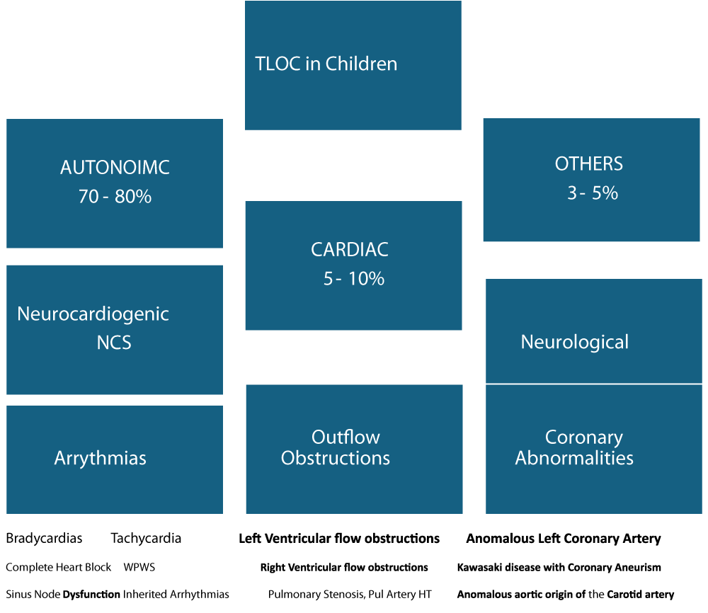
Temporary Loss of Consciousness (TLOC), or syncope, in children is caused by a temporary drop in the blood flowing to the brain. The underlying event in all types of syncope is transient cerebral hypoperfusion [1-4]. Syncope is a common clinical problem in the pediatric age group with most estimates quoting that 15 % of the population would have experienced at least one episode by the age of 18 years [1]. A clinical approach to recognize and treatment of syncope aims to identify the causes as: 1) Autonomic Syncope 2) Cardiac Syncope and 3) Others including neurological causes, based on the underlying risk and pathophysiology [1,4]. Distinguishing between syncopal and non-syncopal TLOC like seizures and elucidating their pathophysiology is crucial for managing syncope. Most of the syncope episode in pediatric age are attributed to autonomic instability, and they are usually benign. However, a small but significant group of children (5% - 10%) may experience symptoms due to a cardiac cause. The differentiation between the two is usually possible by a detailed history and physical examination.
In children i) Situational syncope is a common type of reflex syncope that is associated with typical triggers emotional stress, pain, prolonged standing, dehydration, or the sight of blood, micturition, swallow, cough and hair grooming. ii) This is followed by Vasovagal syndrome a sudden drop in blood pressure with or without a decrease in heart rate, caused by overstimulation of the nerves that have direct input on the heart and blood vessels iii) Rarely Arrhythmia, a heart rate that is too slow, or too fast, or too irregular to keep enough blood flow to the body, including the brain, and iv) Structural (muscle or valvular defects) heart disease, v) Myocarditis an Inflammation of the heart muscle, can also cause fainting [2,4].
In managing syncope in children at primary care, focus is on a thorough history and physical exam, First Aid during an episode, basic investigations of ECG, urgent referral, if need be. Locally managing only if urgent referral is not required includes educating parents, increasing fluid & salt intake, and behavior modification in the longtime. If these measures don’t solve the problem, medications. Referral again if these measures fail to specialists.
This article is a series of anecdotal case reports accompanied by a literature review. It explores the challenges, opportunities, and current evidence in creating a thoughtful diagnostic and management plan for a child or an adolescent with functional neurologic symptom disorder and comorbid cardiac disease. It captures the recent epidemiology, screening, diagnostic, and treatment measures utilized in pediatric syncope with a focus on differentiating psychogenic causes from serious cardiac and benign etiologies. It also describes how psychiatric and psychological factors influence assessment, management, and outcomes.
Case reports
Dental associated syncope
A 4-y-old girl presented with a history of 2–3 episodes per week of syncope-presyncope during the past 4 months in December 2024. She had 4 such episodes prior to this consultation. History revealed that Syncope occurred at “Anganwadi” center and at home when facing verbal or visual stimuli of milk-tooth fall or small wounds in oral cavity. There was nothing remarkable in her personal history, physical examination or studies. During a Tilt-table test she presented with 42 s of asystole with generalized hypertonia and spontaneous urination when a Pediatrician described a dental extraction for the first time. Basic Cardiopulmonary Resuscitation (CPR) was started and the patient made a complete recovery. She was diagnosed with Vasovagal Syncope (VVS) associated with dental phobia and was referred to Child and Adolescent Psychiatry (CAP). After 16 sessions of Cognitive Behavioral Therapy (CBT) and administration of 0.5 mg lorazepam 1 h prior to each session. A continued follow-up with the Cardiologist and by the age 7 years when she lost all her teeth with no syncope symptom.
Fluid restriction induced orthostatic syncope
A 10-year-old male school judo player cum Athlete was brought to the clinic, with the complaints of occasional light headedness after 30 minutes of participation in judo or running race. He reported frequent palpitations during exercise which last for a few minutes before gradually subsiding. He has had two syncopal episodes in the past, one of which occurred while standing and another during judo practice. His pre-Judo or running schedule revealed that he had been following a regimen of extreme fluid restriction several days prior to the competition to maintain his weight class. As physical examination revealed no cause of presyncope. Therefore, he was referred to cardiology check-up. Investigation of electrocardiogram (ECG), 2D Echo (transthoracic echocardiogram) were normal. In a treadmill stress test, he was able to exercise for 10 minutes and 58 seconds, before terminating early due to complaint of dizziness. Heart rate ranged from baseline of 72 beats per minute (bpm) to a maximum of 195 bpm. Baseline blood pressure was 115/54 mmHg and progressively dropped to a low of 94/50 mmHg eleventh minute into the test, rising to 107/56 mmHg after recovery for 10 minutes. When the stress test was performed, the patient had been on a fluid restriction regimen a day prior to preparing for the competition 3 days down the line. He was advised to increase his fluid and salt intake. He has been asymptomatic following increase in fluid intake.
Stretch (Vasovagal) syncope
A 17-year-old male presented for the evaluation of syncope. He had been having syncopal episodes approximately once per month for the past two years, and these episodes had recently increased in frequency. According to the patient’s mother, the episodes were always preceded by arm stretching, while reaching for something from the closet. On inquiry the patient himself reported holding his breath while stretching. During the most recent episode, he was stretching, became dizzy, and subsequently lost consciousness, followed by brief upper extremity convulsions. There was no history of palpitations, shortness of breath, or chest pain. Cardiac evaluation was unremarkable with normal ECG and echocardiogram. The patient was discharged with the impression of vasovagal syncope caused by unintentional Valsalva manoeuvre during stretching, and a subsequent tilt-table test was negative.
Exacerbation of allergy led syncope
A 13-year-old female with a history of asthma and eczema was brought to our clinic with a complaint of syncopal episodes. History revealed that she would get exercise-induced generalized urticaria prior to losing consciousness. A few hours before the episodes, she had flushing, dizziness, rhinorrhea, and shortness of breath. As part of diagnostic workup, an ECG, echocardiogram, cardiac Computerized Tomography (CT), and a cardiac Magnetic Resonance Imaging (MRI), were done and all were normal. However, a treadmill stress test lasted for 11 minutes, with a blood pressure of 142/68 mmHg and heart rate of 151 bpm ten minutes into exercise, and a subsequent drop to a blood pressure of 120/60 mmHg and heart rate of 200 bpm, and was discontinued due to experiencing dyspnea, chest tightness, and generalized urticaria. She was put on antihistamines that resulted in resolution of the episodes.
Pulmonary Arteriovenous Malformation (PAVF) causing Syncope
A ten-month-old female child was brought to me with complaints of bluish discoloration (cyanosis) since birth, tachypnea and failure to thrive. She had developed high grade fever with seizures at 7 months of age. On examination central cyanosis and clubbing was observed, cardiovascular system was normal and there was no murmur in the chest. Her hemoglobin was 14 g/dl and TLC was 6,700/cu mm. ECG showed a heart rate of 110/min, a QRS axis of -60, with no chamber hypertrophy. Chest X-ray revealed a cardiothoracic ratio of 60%. There was a 2.5 x 2.2 cm circular shadow seen in the right lower zone and nonhomogeneous nodular opacities in the right middle & lower zones.
The case was referred to a private tertiary care facility where an Echocardiography revealed a structurally normal heart. Agitated saline, injected as contrast material in the left arm appeared in the left atrium after three cycles, suggestive of a Pulmonary Arteriovenous Fistula (PAVF). Cardiac catheterization revealed normal pulmonary arterial pressures (mean 17 mm Hg) and systemic arterial desaturation (oxygen saturation 66%). Selective pulmonary arterial angiogram delineated multiple large Pulmonary Arteriovenous Malformation (PAVF) arising from a hypertrophied branch of the right pulmonary artery, while the left pulmonary artery was normal. The lower lobe branch fed the largest AVF. Case was diagnosed as PAVF.
PAVF is a rare malformation in a child presenting with cyanosis in the presence of a normal cardiac examination. The patients may have dyspnea, clubbing and clinical evidence of paradoxical embolization and may present as brain abscesses in rare cases. Computed tomography of the brain had revealed bilateral infarcts.
Discussions
Syncope is defined as “a transient loss of consciousness associated with an inability to maintain postural tone with rapid and spontaneous recovery [3]. Pediatric syncope is common, with as many as 50% of all children experiencing at least 1 syncopal event by age 18 [1-3]. Pediatric syncope accounts for 1% - 3% of all emergency department visits and demonstrates a slight female preponderance [2, 3]. Although life-threatening cardiac causes are rare, syncopal and other unresponsive events are distressing for patients, families, and providers.
Neurally Mediated Syncope (NMS)
NMS is the most common form of fainting and a frequent reason for emergency visits by the parents with affected child. It’s also called reflex, neurocardiogenic (NCP), vasovagal (VVS) or vasodepressor syncope (VDS). It’s harmless and rarely requires medical treatment. It is more common in children and young adults, though it can occur at any age. The mechanism involves increased vagal stimulation leading to slowing of heart rate, peripheral vasodilatation, causing hypotension leading to decreased cerebral perfusion. It happens when the part of the nervous system that regulates blood pressure and heart rate malfunctions in response to a trigger, such as emotional stress or pain. It usually happens after standing for a long time, often preceded by a sensation of warmth, nausea, light-headedness, tunnel vision or visual “grey out.” Placing the patient in a supine position helps restore cerebral perfusion and consciousness. Situational syncope, is a type of NMS, related to certain physical functions, like violent coughing, laughing, swallowing or urination. Autonomic Syncope accounts for close to 80% of pediatric cases with TLOC. This pathophysiology allowed clinicians to categorize the phenomenon as the ‘Neuro-Cardiogenic Syncope (NCS)’ also referred to colloquially as “common faint”. It exhibits three components i) a prodrome which almost always precedes ii) the loss of consciousness, followed by iii) a prompt and usually complete recovery. The pathophysiology of NCS is best explained as a paradoxical reflex where pooling of blood in the veins results in both a catecholaminergic surge as well as increased vagal tone [4] known as Bezold-Jarisch reflex. Apart from these 3 components, a careful history will also reveal the presence of precipitants, like decreased individual threshold for the event that triggers the event. The common precipitants in children include hunger, lack of sleep, dehydration, anemia and viral illnesses while typical triggers include sudden change of posture, prolonged upright posture and emotional stress.
Syncope occurring in the supine position suggests a non-autonomic etiology a non-autonomic cause and must be investigated further.
Cardiovascular Syncope (CVS)
CVS is caused by various heart conditions, such as bradycardia, tachycardia or certain types of low blood pressure. It can indicate an increased risk of sudden cardiac death. People suspected of having cardiac syncope but who don’t have serious medical conditions may be managed as outpatients. Further in-patient evaluation is needed if serious medical conditions are present. “Abnormal heart rhythms, coronary artery disease, severe aortic stenosis, and pulmonary embolism are some of the conditions that warrant hospital evaluation and treatment. These conditions can present with various symptoms, including chest pain, shortness of breath, dizziness, or fainting. If an evaluation suggests cardiac or vascular abnormalities, or if you have experienced multiple instances of fainting due to heart-related issues, an ambulatory external or implantable cardiac monitor may be required for continuous monitoring and accurate diagnosis.
Sick sinus syndrome, atrial fibrillation and other serious cardiac conditions can cause recurrent syncope in older adults, rarely seen in children after exercise or exertion, associated with palpitations or irregularities of the heart and usually linked to family history of recurrent syncope, heart disease at a young age or sudden death.
Neuropsychiatric causes of syncope include epilepsy, Functional Neurologic Symptom Disorder (FND) or conversion disorder, psychogenic non-epileptic seizures (PNES), and Psychogenic Non-Syncopal Collapse (PNSC). These conditions account for approximately 10% - 30% of the remaining cases of syncope [3] (Figure 1, Table 1).
The clinicians understanding and perceptions of the causes of Syncope are both a value-add and sometimes a pitfall in the diagnosis and management.
A Tilt Table Test (TTT)
- A TTT also known as an upright tilt test, is a medical procedure used to diagnose syncope (fainting) or other conditions related to the autonomic nervous system, by observing how the body responds to changes in position from lying to standing. It is used to assUnexplained Fainting,
- The function of the autonomic nervous system, which controls heart rate and blood pressure,
- Postural Orthostatic Tachycardia Syndrome (POTS) is a condition characterized by an abnormally high heart rate when standing and
- Other Conditions such as vasovagal syncope and orthostatic hypotension.
Procedure: The patient lies on a special table that can be tilted to an upright position. Healthcare professionals monitor the patient’s blood pressure, heart rate, and other vital signs during the test. The test lasts for 20 - 45 minutes, to see if symptoms like dizziness, fainting, or changes in vital signs occur. In some cases, nitroglycerin or isoproterenol is administered during the test to further assess the autonomic nervous system’s response.
A single private hospital records review, including clinical and laboratory details of children presenting with real or apparent syncope. Five diagnostic categories were identified: Neurocardiogenic Syncope, (NCS), psychogenic pseudo syncope (PPS), cardiac, neurological and indeterminate. Of the 30 children (aged 4 to 17 years) the commonest cause was NCS (63.3%), followed by PPS (13.3%), cardiac (10%), neurological (10%) & indeterminate (3.3%). Exercise, loud noise or emotional triggers and family history were associated with cardiac etiology, and electrocardiogram (ECG) was diagnostic in the majority. Children with PPS and cardiac syncope had frequent episodes when compared with other groups. Indiscriminate antiepileptic use was found in 5 children, including two cardiac cases. Study concluded that frequent recurrences of syncope may suggest PPS or cardiac cause. Cardiac etiology may be identified through clinical history and ECG alone [6].
Out of the 40 patients of syncope 65% were above the age of 10 years with male preponderance (60%). Vasovagal syncope (57%) was the most common cause of syncope followed by orthostatic hypotension (15%), neurological (15%), and cardiac etiology (6%). In the neurological etiology the EEG showed diffuse slow background with occasional sharp bursts in right frontal area in 2 patients while in 4 patients sharp bursts were present in the centero-temporal region. 17% were classified as presyncope, 60% as mild and 22% as having severe syncope. There was a significant correlation of etiology of syncope with duration of hospitalization of more than 4 days and with recurrence of syncope with cardiogenic syncope. On follow up, neurological syncope patients had significant decrease in the number of syncopal episodes as they were immediately started on antiepileptic medicines. This study Inferred that Electrocardiogram, 2D Echocardiogram and Electroencephalogram are important tools for the early management and treatment of cardiac and neurological etiology of syncope [7].
Managing syncope care in primary health care settings
In managing syncope (fainting) in children at primary care, focus is on a thorough history and physical exam, basic investigations, timely referrals, reassurance, and, when referral is not needed, management through parental education, increasing fluid and salt intake, and behavior modification. If these measures don’t solve the problem, medications may be prescribed; if these are ineffective, referral to specialists is reconsidered.
Detailed steps:
- Initial assessment and management: A detailed history focusing on the nature of the syncope (prodromes, triggers, duration, associated symptoms), family history (especially cardiac), and a thorough physical exam are crucial to determine the underlying cause. Both the child and their parents should be reassured that syncope is often a benign condition, & counsel about the common causes and preventative measures. Syncope must be differentiated from seizures, hypoglycemia, and psychiatric disorders.
- First aid during an episode: i) Encourage the child to lie down or sit down with their head between their knees to increase blood flow to the brain ii) While lying, elevate the child’s legs slightly iii) Loosen tight clothing around the neck or waist iv) Allow the child to remain in place until fully alert and reoriented. Medications are usually used as an adjunct therapy, if conservative measure listed above failed. Beta-blockers are used in certain cases of cardiac syncope. Fludrocortisone (e.g., Floricot, Tyroflu) or midodrine (e.g., Gutron 2.5 mg) is used to increase blood volume and improve blood pressure. Additional pharmacological therapies may be considered depending on the underlying etiology.
- Urgent referrals: If there is a strong clinical suspicion of a cardiac etiology, such as a family history of sudden cardiac death, syncope during exercise, heart murmur, or abnormal ECG), refer to a pediatric cardiologist for further evaluation. Similarly, children with frequent or prolonged syncopal episodes must be referred for cardiac & neurological assessments. If the cause of syncope remains unclear after a thorough evaluation, referral to a specialist be considered after primary treatment & counselling. If syncope is accompanied by neurological symptoms like seizures, prolonged loss of consciousness, such cases also must be referred to a pediatric neurologist.
- Managing at primary care setting: Once the need for urgent referral is ruled out i) Encourage adequate hydration and salt intake, particularly during hot weather or strenuous activity ii) Advise the child to sit or lie down when feeling lightheaded, stand up slowly, and avoid prolonged standing or dehydration iii) Identify and avoid situations or activities that seem to precipitate syncope.
- Psychological well-being: As recurrent syncope can have a negative impact on a child’s psychological well-being, any anxiety or fear associated with fainting should be addressed proactively. Long-term management includes lifestyle adjustments, appropriate pharmacotherapy, and regular follow-up, medications, and regular follow-up with a healthcare provider.
Conclusion
Syncope is common among children and adolescents, most often benign, but can significantly impact quality of life, contributing to psychiatric comorbidities. In pediatric patients with recurrent syncope or comorbid cardiac disorders, the importance of performing a careful history and physical exam is foundation for the diagnostic assessment. Differentiating between aetiologies of syncope is dependent on characterizing the situation in which syncope occurred, prodromal symptoms, semiology. In managing syncope in children at primary care, urgent referral if required and if not at managing at the primary care level by increasing fluid and salt intake, reassurance, to the child and the parents, medications and behavior modification. If these measures are ineffective, referral to specialists or advanced therapies must be considered.
- Krishna MR, Kunde MF. A clinical approach to syncope. Indian J Pract Pediatr. 2020;22(1):92. Available from: https://www.ijpp.in/Files/2020/ver1/A%20clinical%20approach%20to%20syncope.pdf
- Yeom JS, Woo HO. Pediatric syncope: pearls and pitfalls in history taking. Clin Exp Pediatr. 2023;66(3):88–97. Available from: https://doi.org/10.3345/cep.2022.00451
- Lukich SD, Sarin A, Pierce JM, Russell MW, Malas N. Syncope and unresponsiveness in an adolescent with comorbid cardiac disease: an illustrative case report and literature review of functional neurologic symptom disorder. J Acad Consult Liaison Psychiatry. 2023;64(4):392–402. Available from: https://doi.org/10.1016/j.jaclp.2023.03.006
- Brignole M, Moya A, de Lange FJ, Deharo JC, Elliott PM, Fanciulli A, et al. 2018 ESC Guidelines for the diagnosis and management of syncope. Eur Heart J. 2018;39(21):1883–1948. Available from: https://doi.org/10.1093/eurheartj/ehy037
- Grubb BP. Clinical practice: neurocardiogenic syncope. N Engl J Med. 2005;352(10):1004–10. Available from: https://doi.org/10.1056/nejmcp042601
- Mohanty S, Kumar CPR, Kaku SM. Clinico-etiological profile of paediatric syncope: a single centre experience. Indian Pediatr. 2021 Feb 15;58:132–4. Available from: https://pubmed.ncbi.nlm.nih.gov/33632942/
- Fadnis M, Prabhu S, Venkatesh S, Kulkarni S. Syncope in children: clinic-aetiological correlation. Int J Contemp Pediatr. Available from: https://www.ijpediatrics.com/index.php/ijcp/article/view/2854
Article Alerts
Subscribe to our articles alerts and stay tuned.
 This work is licensed under a Creative Commons Attribution 4.0 International License.


 Save to Mendeley
