Global Journal of Rare Diseases
Ortner’s Syndrome in the Modern Era: A Series of 7 Cases
AKM Monwarul Islam1*, Ishrat Jahan Shimu2, Kaniz Fatema Ananya2, Rezvey Sultana2 and Saima Alam3
2Assistant Registrar, Department of Cardiology, National Institute of Cardiovascular Diseases, Dhaka, Bangladesh
3Honorary Medical Officer, Department of Cardiology, National Institute of Cardiovascular Diseases, Dhaka, Bangladesh
Cite this as
Monwarul Islam AKM, Shimu IJ, Ananya KF, Sultana R, Alam S (2018) Ortner’s Syndrome in the Modern Era: A Series of 7 Cases. Glob J Rare Dis 3(1): 001-004. DOI: 10.17352/2640-7876.000010Ortner syndrome (OS) is a rare condition characterized by hoarseness of voice in association with a cardiovascular disease. Mitral stenosis is a well-recognized cause; however, because of control of rheumatic fever and rheumatic heart disease in many parts of the world, there may be a shift of the underlying aetiology to other cardiac and non-cardiac conditions. Aortic diseases and pericardial effusion as the cause of OS, are being increasingly recognized. Here, 7 cases of OS have been presented, with special attention to their clinical presentation, underlying pathology, diagnostic work-up, treatment offered and the prognosis observed. Three cases expired, 2 cases were managed successfully, while 2 cases are waiting for surgery. In all cases, hoarseness of voice did not improve. OS should be a differential diagnosis while dealing with a patient with otherwise unexplained hoarseness of voice.
Introduction
Ortner syndrome (OS), also called cardiovocal syndrome, refers to left recurrent laryngeal nerve palsy due to cardiovascular disease. Classically, this was reported with dilated left atrium due to mitral stenosis [1]. However, subsequently, a number of non-valvular conditions have been found to cause OS [2]. The prevalence of OS is unknown, presumably due to rarity of the condition. Also, because of control of rheumatic heart disease in many parts of the world, the aetiopathological aspect of OS has likely to be changed. Here, we describe a series of OS cases documented over the last 7 years between 2011 and 2018 (Table 1). The clinical profile of the patients including follow up has been presented.
Case Presentation
Case 1
A 35-year-old lady [3], presented with progressive breathlessness, recurrent episodes of productive cough and occasional haemoptysis for 6 months, and hoarseness of voice for 1 month. She was on anti-TB drugs for the presumptive diagnosis of pulmonary tuberculosis. Echocardiography revealed moderate mitral stenosis, only mildly dilated left atrium (41 mm) and severe pulmonary hypertension (pulmonary artery systolic pressure 92 mmHg). High resolution CT scan of the chest revealed features of bilateral bronchiectasis, whereas pulmonary arteriography found dilated pulmonary arteries. Left vocal cord palsy was detected in fibreoptic laryngoscopy (FOL). She was given rheumatic fever prophylaxis, additional antibiotics, chest physiotherapy, bronchodilators, diuretics and nifedipine. Over the next months, there was gradual improvement of the condition of the patient except the hoarseness of voice. While staying in the village, suddenly her condition deteriorated and on the way to the hospital she expired.
A 35-year-old man [4], presented with low-grade fever, cough and occasional haemoptysis for 3 months and hoarseness of voice for 1 month. Physical examination was unremarkable. His investigations revealed: ESR 110 mm in first hour, Mantoux test positive (15 mm), acid fast bacilli in bronchoalveolar lavage fluid, left hilar lesion in chest X-ray, and left vocal cord palsy in FOL. He was offered anti-TB drugs, but while on anti-TB therapy, the patient developed huge pericardial effusion. Pericardiocentesis was done; the cultured TB bacilli revealed resistance to rifampicin, but sensitivity to INH, ethambutol and streptomycin. The patient was treated conservatively with INH, ethambutol, pyrazinamide, streptomycin and ofloxacin. Over the next months, there was little improvement, pericardial fluid reaccumulated, and the hoarseness of voice did not improve significantly. Meanwhile, left-sided pleural effusion developed, pleural biopsy abroad reported presence of suspicious malignant cells. Fine-needle aspiration cytology (FNAC) of cervical lymph nodes established the diagnosis of metastatic adenocarcinoma. The patient was offered 6 cycles of palliative chemotherapy. Cisplatin was instilled intrapericardially and intrapleurally to prevent recurrence. General condition of the patient improved significantly, and there was almost no fluid in the pericardial and pleural spaces in follow-up. However, about 2 years after presentation, he ultimately, succumbed to death.
Case 3
A 30-year-old man presented with dyspnoea and dysphagia for 6 months, and hoarseness of voice for 6 weeks. His investigations revealed: atrial fibrillation in ECG, and mitral valve prolapse, severe mitral regurgitation, dilated left atrium (58 mm) and moderately depressed left ventricular ejection fraction (LVEF 30-35%) in echocardiography, and left vocal cord palsy in FOL. He was offered conservative treatment with diuretics, angiotensin-converting enzyme inhibitor and digoxin. He refused any surgical treatment. There was clinical improvement, however, the hoarseness of voice did not change significantly. Two years after presentation, he is alive.
Case 4
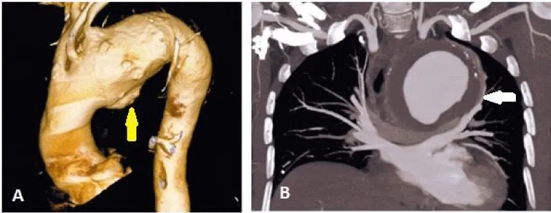
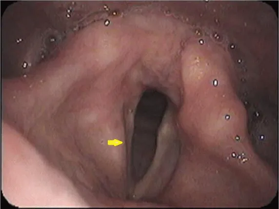
A 58-year-old man presented with chest pain, vertigo and hoarseness of voice. His investigations revealed: suspected aortic aneurysm in chest x-ray, dilated aortic root and ascending aorta, normal-sized left atrium (27 mm) in echocardiography, large aneurysm of arch of aorta, mural thrombus and dissection in CT aortogram (Figure 1), and left vocal cord palsy in FOL (Figure 2). He was offered conservative treatment with beta-blocker, and was referred for surgery. After successful surgical treatment of aortic aneurysm, he is doing well, but the hoarseness of voice did not change.
Case 5
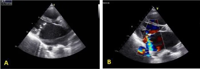
A 29-year-old man presented with effort intolerance, cough, and hoarseness of voice. His investigations revealed: right ventricular hypertrophy, biatrial enlargement and sinus rhythm in ECG, and severe mitral stenosis (MVA 0.7 cm2), severe mitral regurgitation, dilated left atrium (59 mm) and severe pulmonary hypertension (PASP 90 mmHg) in echocardiography (Figure 3). FOL revealed left vocal cord palsy. He was given conservative treatment, and was sent for surgery. However, after 4 months, while waiting for mitral valve replacement, he expired.
Case 6
A 64-year-old man presented with dyspnoea, heaviness in the chest and hoarseness of voice. His investigations revealed: cardiomegaly in chest X-ray, and huge pericardial effusion in echocardiography. Pericardiocentesis revealed haemorrhagic fluid, exudative in nature, adenosine deaminase (ADA) was raised (62.84 U/l), gene Xpert test was negative for Mycobacterium tuberculosis, while FOL revealed left vocal cord palsy. He was given anti-TB treatment, and the patient is doing well now.
Case 7
A 38-year-old man presented with dyspnoea, palpitation and hoarseness of voice. His investigations revealed: atrial fibrillation in ECG; severe mitral stenosis, mild mitral regurgitation, moderate aortic regurgitation, dilated left atrium (52 mm) and PASP 72 mmHg in echocardiography. FOL revealed left vocal cord palsy. He was given conservative treatment, however, hoarseness of voice did not improve significantly. He is waiting for double valve replacement.
Discussion
OS is an uncommon condition. The 7 cases presented here have been documented over 7 years between 2011 and 2018. However, because of rarity and lack of proper attention to changes in voice in a patient with cardiovascular or other pathology, this is likely that some cases might be missed. OS was first described by Nobert Ortner, a Viennese physician in 1897 [1], and subsequently, by others, in cases of mitral stenosis [5-7] In our series, mitral stenosis was the underlying cause in 3 out of 7 cases, however, in 1 case [3] mitral stenosis was complicated by bronchiectasis causing pulmonary hypertension. Another case of OS was due to mitral valve prolapse accompanied by severe mitral regurgitation and hugely dilated left atrium. Kishan et al. described OS in an 83-year-old woman with MVP [8]. Pericardial effusion appears to be an important cause, and it was found in 2 cases presented here. In 1 case, tuberculosis was confirmed, whereas in another case, dual pathology was likely, i.e., tuberculosis and malignancy [4]. Tuberculosis is a common cause of pericardial effusion in South Asia, so, this infectious disease may be an important cause of OS regionally. Aortic aneurysm as a cause of OS was reported well previously [9-12]. In our series, we found aneurysm of the arch of the aorta in 1 case. So, a myriad of valvular and non-valvular conditions may, in fact, cause this cardiovocal syndrome. This has also been shown in a collective review where aortic disease was the commonest cause of OS [2].
Left recurrent laryngeal nerve palsy causes the hoarseness in OS. The nerve injury is commonly caused by the pressure effect on the nerve by the dilated left atrium in mitral stenosis. Later, it was apparent that, other structures like dilated pulmonary artery, aortic aneurysm or even pericardial effusion may also exert the pressure effect on the nerve. In fact, the current understanding favours pressure in the pulmonary artery playing the most important role in causing the nerve compression in a majority of the cases [13]. In our observation, left atrium was, in fact, of normal diameter (<40 mm) in 3 cases, and only marginally dilated (41 mm) in 1 case. On the other hand, pulmonary artery systolic pressure was significantly elevated in 3 of our cases.
In OS, the underlying conditions are acquired ones, lacking any genetic predisposition. In our series, none of the family members had similar type of illness. 0n the other hand, high degree of clinical suspicion is needed for making the diagnosis of OS. Availability of FOL has eased the confirmation of vocal cord palsy, as is the issue in our series.
Treatment of OS is mainly directed to the correction of underlying pathology which may not always be easy. Three of our patients expired within 2 years of diagnosis of OS, 1 patient improved with treatment of tubercular pericardial effusion, 1 patient is doing well after surgery of aortic aneurysm, while 2 patients are still waiting for surgery. However, during the period of observation, hoarseness of voice did not improve in any patient. The explanation behind this gloomy prognosis of hoarseness may be late diagnosis causing permanent injury to the nerve. Early management of underlying condition and advanced treatment of vocal cord palsy might improve hoarseness of voice.
OS is a rare entity. Though mitral stenosis is the classical cause, other cardiac and non-cardiac causes are being increasingly recognized. Tuberculosis may be an important aetiology in endemic zone. High degree of clinical suspicion is essential for making the diagnosis. Hoarseness of voice may be irreversible by the time the diagnosis is made. Prognosis is mainly determined by underlying condition. Even in this era, OS may be associated with high mortality.
Author’s Contributions
AKM Monwarul Islam contributed for the diagnosis and management of the cases and also drafted the manuscript. Ishrat Jahan Shimu, Kaniz Fatema Ananya, Rizvi Ahmed and Saima Alam were associated with diagnosis and management of the patients, and provided intellectual thoughts to the manuscript.
- Ortner N (1897) Recurrent laryngeal nerve paralysis due to mitral valve stenosis. Wien Klin Wochenschr 10: 753-755.
- Yuan S (2014) Ortner (cardio-vocal) syndrome: A collective review. Kuwait Medical Journal 46: 3-13. Link: https://goo.gl/EY76K1
- Monwarul Islam AKM, Zaman S, Doza F (2012) Ortner syndrome due to concomitant mitral stenosis and bronchiectasis. Korean Circ J 42: 507-510. Link: https://goo.gl/LG9EUK
- Islam AKMM, Rahman MT, Ali M (2013) Ortner syndrome with recurrent pericardial effusion: a diagnostic and therapeutic dilemma. J Coll Physicians Surg Pak. 23: 660-662. Link: https://goo.gl/292ca5
- Subramaniam V, Herle A, Mohammed N, Thahir M (2011) Ortner's syndrome: case series and literature review. Braz J Otorhinolaryngol 77: 559-562. Link: https://goo.gl/9bTUHd
- Plastiras SC, Pamboucas C, Zafiriou T, Lazaris N, Toumanidis S (2010) Ortner’s syndrome: a multifactorial cardiovocal syndrome. Clin Cardiol 33: E99-100. Link: https://goo.gl/e61BV6
- Mohamed AL, Zain MM (2004) Hoarseness of voice in a patient with mitral stenosis and Ortner's syndrome. Malays J Med Sci 11: 65-68. Link: https://goo.gl/nFnxwe
- Kishan CV, Wongpraparut N, Adeleke K, Frechie P, Kotler MN (2000) Ortner's syndrome in association with mitral valve prolapse. Clin Cardiol 23: 295-297. Link: https://goo.gl/hu7vmL
- Kyaw WA, Lim CY, Khalil MAM, Lim KC, Chong VH, et al. (2018) A fatal case of Ortner's syndrome and dysphagia aortica secondary to rapidly expanding mycotic thoracic aortic aneurysm in a chronic kidney disease patient. SAGE Open Med Case Rep 6: 2050313X18799247 Link: https://goo.gl/3RQmVh
- Semionov A, Kosiuk J (2016) Ortner syndrome secondary to aortic aneurysm. Radiol Case Rep 12: 29-30. Link: https://goo.gl/3zdBRx
- Ismazizi Z, Zainal AA (2016) Thoracic aortic aneurysm as a cause of Ortner's syndrome - A case series. Med J Malaysia 71: 139-141. Link: https://goo.gl/AfpBKS
- Escribano JF, Carnés J, Crespo MA, Antón RF (2006) Ortner’s syndrome and endoluminal treatment of a thoracic aortic aneurysm: a case report. Vasc Endovascular Surg 40: 75-78. Link: https://goo.gl/ByThHc
- Lee SI, Pyun SB, Jang DH (2006) Dysphagia and hoarseness associated with painless aortic dissection: a rare case of cardiovocal syndrome. Dysphagia 21: 129-132. Link: https://goo.gl/923iGL

Article Alerts
Subscribe to our articles alerts and stay tuned.
 This work is licensed under a Creative Commons Attribution 4.0 International License.


 Save to Mendeley
