Global Journal of Rare Diseases
Idiopathic Gingival Elephantiasis – A Case Report
Nasim Aarfa1, Sasankoti Mohan Ravi Prakash2*, Malik S Sangeeta3 and Gupta Swati4
2Professor & Head, Oral Medicine and Radiology, Subharti Dental College, Meerut, India
3Reader, Oral Medicine and Radiology, Subharti Dental College, Meerut, India
4Reader, Oral Medicine and Radiology, Subharti Dental College, Meerut, India
Cite this as
Aarfa N, Ravi Prakash SM, Sangeeta MS, Swati G (2017) Idiopathic Gingival Elephantiasis – A Case Report. Glob J Rare Dis 2(1): 015-017. DOI: 10.17352/2640-7876.000009Gingival elephantiasis is a rare slow progressive lesion which is also known as gingival fibromatosis. It can be localized or generalized. This condition can be inflammatory, non-inflammatory or combination of both. The etiology involved could be due to poor oral hygiene, inadequate nutrition, a systemic hormonal stimulation. Genetic cause has been implicated as its etiology with several genes mutations and sometimes associated with syndromes such as Cross syndrome, Rutherford syndrome or Ramen syndrome but isolated cases are also reported and their etiology often remains unknown. This paper reports a very interesting case in which a 35 year old female came with complain of generalized swelling of gingiva covering the occlusal surface with mobility of teeth. Patient was asymptomatic and there was progressive increase in growth of gingiva since 1 year. Histopathological diagnosis of gingival growth confirmed the diagnosis.
Introduction
Elephantiasis gingivae is also known as fibromatosis gingivae, gingivostomatitis, hereditary gingival fibromatosis, idiopathic fibromatosis, familial elephantiasis, and diffuse fibroma [1]. It is defined as a rare, benign, asymptomatic, nonhemorrhagic and nonexudative proliferative fibrous lesion of the gingival tissues [1]. It was first reported by Goddard and Gross in 1856 [2]. It is characterized by a slowly progressive, benign enlargement, which affects mainly the marginal gingiva, attached gingiva, and interdental papilla. The fibromatosis may potentially cover the exposed tooth surfaces, causing esthetic and functional problems, and in some cases distort the jaws. Gingival tissues surrounding both the maxillary dentition and the mandibular dentition may be affected. The hyperplasic gingiva usually presents a normal color and has a firm consistent with abundant stippling [3]. Men and women are equally affected at a phenotypic frequency of 1:750 000 with varying intensity and expressivity even in individuals within the same family [4-7]. Hereditary Gingival Fibromatosis may be associated with other clinical manifestations such as hypertrichosis, hypopigmentation, mental deficiency, epilepsy, splenomegaly, optic and auditory defects, cartilage and nail defects and Dentigerous cyst. It manifests as an autosomal dominant or less commonly, an autosomal recessive mode of inheritance [8,9]. Autosomal-dominant forms of gingival fibromatosis, which are usually nonsyndromic, have been genetically linked to the chromosomes 2p21-p22 and 5q13-q22 [10]. In modern era, a mutation in the Son of sevenless-1(SOS-1) gene has been suggested as a possible cause of isolated (nonsyndromic) gingival fibromatosis. However, no definite linkage has been established [4].
This anomaly is classified into two types according to its form—the nodular form and the symmetric form. The localized nodular form is characterized by the presence of multiple enlargements in the gingiva. The symmetric form which is the most common type of this disorder results in uniform enlargement of the gingiva. The degree of enlargement may vary from mild to severe. As a result, the teeth become buried, to varying degrees, beneath the redundant hyperplastic tissues. The gingival tissues are usually pink and non-hemorrhagic and have a firm, fibrotic consistency [11,12]. Diffuse gingival enlargement is also found to be associated with syndromes like Cross syndrome, Rutherford syndrome, Ramen syndrome, Zimmerman Laband syndrome, and Juvenile hyaline syndrome [11-13].
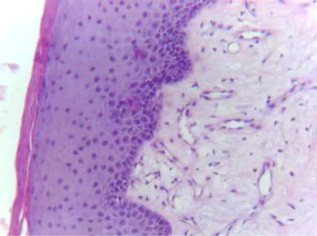
The typical histologic appearance of the affected tissue includes hyperplastic epithelium with elongated rete ridges extending deeply into the underlying connective tissue with coarse and fine dense bundles of collagen, oriented in all directions, and a few “plump” fibroblasts have been described as making up the connective tissue layer.
Case Report
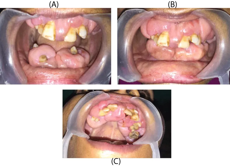
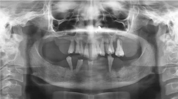
A 35 year old middle aged woman reported to the Department of Oral Medicine and Radiology of our institution with a complaint of swelling of gums in both upper and lower jaw from past 1 year. Patient gave history of bleeding of gums and pain after which swelling started to develop which was causing functional and masticatory difficulty. Patient had undergone extraction and swelling got relieved from that area. There was no past medical and dental history. Patient was afebrile and there was no drug history associated with the disease. On intraoral examination generalized severe gingival overgrowth, involving the maxillary and mandibular arches and covering almost the whole dentition. Enlargement involving both buccal and lingual/palatal sides with pinkish red, fibrous inconsistency and absence of stippling. Gingival enlargement enclosed the major surface of the teeth present engaging the incisal/occlusal surfaces as well. Severe diffuse enlargement involving the marginal, interdental, and attached gingiva of both arches, covering almost all the surfaces of the teeth was found as shown in figure 1 a, b, c. Mobility in all the teeth was present with severe pathologic migration. Radiographical examination in the form of panoramic view revealed severe generalized alveolar bone loss, which could be attributed to the local factors which must have exaggerated the hyperplastic condition. Figure 2 the blood examination results were normal and correlated with an absence of any history of systemic disease. Based on all these findings, a provisional diagnosis of idiopathic gingival elephantiasis was made. The differential diagnosis of the lesion that could be made were Drug induced gingival enlargement, Scurvy, Sarcoidosis, Crohn’s diseases, Cowden’s syndrome, Amyloidosis. Histopathological features revealed highly fibrous connective tissue, with haphazardly arranged dense collagen bundles, numerous spindle shaped fibroblasts and connective tissue that is relatively avascular. Thickened, acanthotic and hyperkeratotic stratified squamous epithelium was also present with elongated rete ridges (figure3).The histopathologic features led to the final diagnosis of idiopathic gingival fibromatosis.
Discussion
Gingival fibromatosis is a rare, benign, non-haemorrhagic fibrous enlargement of gingival tissue. It was previously called elephantiasis gingivae, hereditary gingival hyperplasia and hypertrophic gingiva [14]. It can be congenital or hereditary. Gingival overgrowth varies from mild enlargement of isolated interdental papillae to segmental or uniform and marked enlargement affecting one or both jaws [15]. In the present case, the patient had no history of any systemic disease, hypertrichosis, mental retardation, epilepsy, or medication which could contribute to gingival overgrowth. She also did not give history of pregnancy. General physical examination of the patient revealed no syndromic association which could contribute to gingival overgrowth. The clinical, histopathological features and systemic examination excluded the diagnosis of neoplastic enlargement, hereditary gingival fibromatosis, Wegener’s granulomatosis, acanthosis nigricans [16,17]. The mechanism of idiopathic gingival fibromatosis is unknown, but it is seen often to confine to the fibroblasts which harbor in the gingivae. The hyperplastic response does not involve the periodontal ligament and occurs peripheral to the alveolar bone within attached gingivae [15]. In our case those site where extraction has been done there was no gingival overgrowth present. Authors report this condition as an increase in the proliferation of gingival fibroblasts [18]. Whereas others report slower-than normal growth [19]. Increased collagen synthesis rather than decreased levels of collagenase activity may be involved [18]. In recent studies, hearing loss and supernumerary teeth have been associated with hereditary gingival fibromatosis [20]. The condition has also been reported in association with deficiency of growth hormone caused by lack of growth hormone release factor [21]. The surgical treatment of choice is the gingivectomy, which was first advocated for drug-induced gingival enlargement in 1941. Treatments vary according to the degree of severity of gingival enlargement [14]. When the enlargement is mild, thorough scaling of teeth and proper home care may be sufficient to restore good oral health and appearance. However, if scaling is proved to be ineffective and the gingival overgrowth continues to affect appearance and function, surgical intervention is required. Gingival enlargement with deep pockets and severe loss of underlying alveolar bone, an internal bevel gingivectomy with open flap debridement is required [17]. Flap surgery can be carried out in areas with inadequate attached gingiva [4]. To restore normal gingival appearance and contours gingivoplasty with blades, surgical knives, laser or electro surgery are also the treatment of choice [4]. The recurrence rate is very high so close examination is required with proper maintenance of oral hygiene.Conclusion
We conclude this case as an interesting case where there was excessive gingival overgrowth covering both maxilla and mandibular arches with complete presence of fibrous tissues on all the occlusal surfaces of teeth. This case can be treated as an isolated finding because of its massive proliferative growth of the tissues. Treatment was appreciably done to improve the patient’s aesthetic and masticatory competence as well as her periodontal condition.
- Tavargeri A (2004) Idiopathic gingival fibromatosis—a case report. J Indian Sot Pedo Prev Dent 22: 180-182. Link: https://goo.gl/u17qg1
- Larissa P (2000) hereditary gingival fibromatosis: Review of literature and a case report. Quintessence 31: 415-418. Link: https://goo.gl/YHeBdn
- Bansal A, Narang S, Sowmya K, Sehgal N (2011) Treatment and two year follow-up of a patient with hereditary gingival fibromatosis. J Indian Soc Periodontol 15: 406-409. Link: https://goo.gl/HsWbYu
- Ramakrishnan T, Kaur M (2010) Multispeciality approach in the management of patient with hereditary gingivalfibromatosis:1-year follow up: a case report. Int J Dent. Link: https://goo.gl/WkrQPT
- Cholakis AK, Wiltshire WA, Birek C (2002) Treatment and long term follow-up of a patient with hereditary gingival fibromatosis: a case report. J Can Dent Association 68: 290–294. Link: https://goo.gl/pMfUaX
- Vishnoi SL (2011) hereditary gingivalfibromatosis: report of four generation pedigree. Int J Case Rep Images 2: 1–5. Link: https://goo.gl/mGrNmt
- Zhou M, Xu L, Meng HX (2011) Diagnosis and treatment of a hereditary gingival fibromatosis case. 14: 155–158. Link: https://goo.gl/aFbn38
- Jaju PP, Desai A, Desai RS, Sushma PJ (2009) Idiopathic gingival fibromatosis: case report and its management. Int J Dent 2009:1–6. Link: https://goo.gl/D2ULP5
- Harikihan G (2010) Idiopathic fibromatosis of gingival: a case report of hereditary tendency in the same family. J Indian Acad Dent Spec 1: 52–54.
- Hart TC, Pallos D, Bozzo L (2000) Evidence of genetic heterogeneity for hereditary gingival fibromatosis. Journal of Dental Research, 79: 1758–1764. Link: https://goo.gl/tehegs
- Carranza FA, Hogan EL (2002) Gingival enlargement,” in Clinical Periodontology, M.G. Newman, H.H. Takei, and F.A. Carranza, Eds. 279–296.
- Bakaeen G, Scully C (1998) hereditary gingival fibromatosis in a family with the Zimmermann-Laband syndrome. 20: 457–459. Link: https://goo.gl/ViwqRj
- Gorlin RJ, Pinborg JJ, Cohen MM (1976) Syndromes of the Head and Neck. McGrawHill,
- Mohan RPS, Verma S, Agarwal N (2013) Singh U Non-syndromic hereditary gingival fibromatosis. BMJ Case Rep 10: 1-4. Link: https://goo.gl/19ZjB9
- Tiwana PS, De Kok IJ, Stoker DS, Cooper LF (2005) Facial distortion secondary to idiopathic gingival hyperplasia: surgical management and oral reconstruction with endosseous implants, Oral Surgery, Oral Medicine, Oral Pathology. Oral Radiology and Endodontology 100: 153–157. Link: https://goo.gl/vbam82
- Hart TC, Pallos D, Bozzo L (2000) Evidence of genetic heterogeneity for hereditary gingival fibromatosis. Journal of Dental Research 79: 1758–1764 . Link: https://goo.gl/Tffcbw
- Chaturvedi R (2009) Idiopathic gingival fibromatosis associated with generalized aggressive periodontitis: a case report. Journal of the Canadian Dental Association 75: 291–295. Link: https://goo.gl/5WPkpR
- Shirasuna K, Okura M, Watatani K, Hayashido Y, Saka M, et al. (1988) Abnormal cellular property of fibroblasts from congenital gingival fibromatosis, Journal of Oral Pathology 17: 381–385. Link: https://goo.gl/8TZW6x
- Tipton DA, Howell KJ, Dabbous MK (1997) Increased proliferation, collagen, and fibronectin production by hereditary gingival fibromatosis fibroblasts 68: 524–530. Link: https://goo.gl/aUGUJS
- Oikarinen K, Salo T, Kaar ML, Lahtela P, Altonen M (1990) Hereditary gingival fibromatosis associated with growth hormone deficiency. Br J Oral Maxillofac Surg 28: 335-339. Link: https://goo.gl/B5pWYa
- Wynne SE, Aldred MJ, Bartold MP (1995) hereditary gingival fibromatosis associated with hearing loss and supernumerary teeth -a new syndrome. J Periodontol 66: 75-97. Link: https://goo.gl/KpjbXp

Article Alerts
Subscribe to our articles alerts and stay tuned.
 This work is licensed under a Creative Commons Attribution 4.0 International License.


 Save to Mendeley
